Lightsheet Microscopy: Technology and Advantages
Lightsheet Microscopy: Technology and Advantages
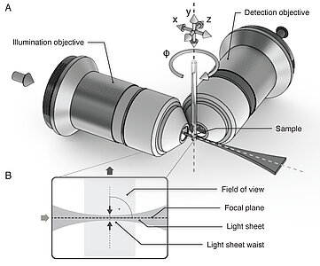
In Light Sheet Fluorescence Microscopy (LSFM), also called Single Plane Illumination Microscopy (SPIM ), illumination and detection are two separate optical systems that are perpendicular to each other. The laser beam used to illuminate the sample is focused in one direction and forms a light sheet that illuminates a thin layer of the sample. The fluorescent light that is emitted in this plane is captured and detected by an objective lens.
The separation of illumination and observation brings two decisive advantages:
- It makes it possible to build up the microscope virtually "around the experiment. Instead of mounting samples in between microscope slide and coverslip, they are immersed in a hydrogel and placed in a cylindrical sample chamber inside the microscope. There, the conditions are close to physiological and so even continuous recordings from living specimens are possible.
- With LSFM, the light exposure of the samples is very low, which considerably reduces phototoxic effects. Only then long-term experiments are possible.
Sample Movement in the Microscope
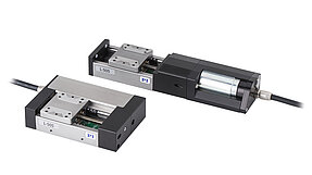
One of the most common application in Light Sheet Microcopy is the creation of Z stacks with which the sample can be wholly depicted in three dimensions. For this purpose, the sample is moved in the Z direction (that is, along the optical axis of the objective lens). For this task, PI contributes compact linear stages to the Flamingo Lightsheet microscopes of the >> Huisken Lab at the Georg-August-University Göttingen. These very compact linear stages have a folded drivetrain, DC motors with gearhead, linear encoders, and offer 26 mm travel range at a bidirectional repeatability of ±250 nm. The stage also excels by a high stiffness and guiding accuracy due to ball guides.
Rotating the specimen can be useful in several scenarios. Often, researchers want to be able to image their sample from just the right angle (side view, top view etc.) or want to image an organ that’s only visible from certain angles (like the heart). The other big application for rotation is so-called multi-view imaging, where z-stacks are recorded from multiple angles, e.g. 6 angles covering 360 degrees, and then the useful parts of each dataset are fused to generate one 3D image that covers the entire sample. This is particularly useful for larger samples as the sample itself scatters, refracts and absorbs the light, affecting the illumination and detection quality of structures deep inside the sample. Therefore several angles are needed to capture all the details.
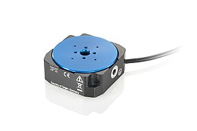
The rotation of the sample is supported by the U-628 ultrasonic piezo-motor-driven rotation stage from PI. It offers fast movement of up to 720°/s at a minimum step size of 51 µrad and a bidirectional repeatability of ±102 µrad.
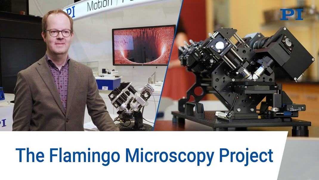
Flamingo LSFM: Compact and Modular Design
A special feature of the concept for the Flamingo light-sheet microscopes is that they are sent as a compact kit to the users. In order for the implementation to work, some conditions had to be met: The system had to be very robust, easy to assemble, and simple to operate. The solution: A straightforward, uncomplicated design with proven standard components, a small footprint, and costs as low as possible. This also allows for different, application-specific designs: In the T-SPIM design, the sample is illuminated from two opposing optics for a more uniform illumination. The addition of a second detection path results in the X-SPIM design.
The Flamingo Project and the resulting microscopes was created by Jan Huisken and his group at the >> Morgridge Institute for Research in Madison, WI. PI is one of the sponsors of the Flamingo Project.
Further information can also be found in our >> blogpost.

